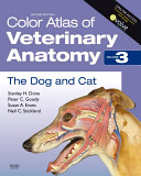
Color Atlas of Veterinary Anatomy Vol. 3. Dog
Atlas pomáhá čtenářům seznámit se s veterinární anatomií. Je obzvláště užitečná během pitevních kurzů k identifikaci struktur a výborná je i při opakování. Každá barevná fotografie je doplněna barevnou přehlednou ilustrací, který skutečně pomůže pochopit, kde se co lokalizuje daná anatomická struktura. Tato publikace rozhodně stojí za vynaložené peníze. Spolu s učebnicí, jako je "Dyceova veterinární anatomie“ jsou základními referenčními zdrojem informací nejen pro studenty, ale i praktické veterináře.
Pes a kočka je třetím svazkem série o veterinární anatomii (přičemž svazky I a II jsou „Přežvýkavci“ a „Kůň“). Důležité rysy topografické anatomie jsou prezentovány v sérii barevných fotografií detailních pitev. Struktury jsou identifikovány v doprovodných barevných kresbách, jejichž cílem je objasnit vztahy mezi příslušnými strukturami. V případě potřeby jsou v popiscích fotografií uvedeny informace potřebné k interpretaci. Kniha začíná fotografiemi regionálních povrchových prvků pořízených před pitvou a v následujících kapitolách je podrobně znázorněna pitva každé části.
Pitvy, fotografie a kresby byly pro tuto knihu speciálně připraveny. Pitvy primárně zobrazují chrta, ale kde je třeba ilustrovat důležité odchylky jsou použity boxera a kočku. Cílem těchto pitev a fotografií je odhalit topografii zvířete tak, jak by byla prezentována veterinárnímu chirurgovi během rutinního klinického vyšetření. Proto převažují boční pohledy. Autoři se vědomě vyhýbají fotografiím částí odebraných z těla nebo použití pohledů z neobvyklých úhlů či neobvyklých tělesných poloh. S touto publikací budou veterinární chirurgové a studenti schopni vidět pod vnějším povrchem zvířat svěřených do jejich péče. Pacientovy svaly, kosti, cévy, nervy a vnitřnosti, které tvoří každou oblast těla a každý orgánový systém.
Autor: John S. Boyd BVMS, PhD. MRCVS, Professor of Veterinary Anatomy, University of Glasgow Veterinary School, UK, Callum Paterson MSc, Reserch Technician, Department of Veterinary Anatomy, University of Glasgow, UK
| Nakladatel | Elsevier Saunders |
|---|---|
| ISBN | 9780723434153 |
| Vydání | 2. vydání 2000 |
| Vazba | brožované |
| Počet stran | 224 |
This book is really helping me to get to grips with veterinary anatomy. It is especially useful during dissection classes to help identify structures, and excellent during revision as well. Each clear photo is accompianied by a colour line diagram which really helps you understand where everything is, and is an advantage over Boyd 'Clinical Anatomy of the Dog and Cat', this book is well worth the extra expense. Together with a good textbook such as Dyce 'Veterinary Anatomy' this provides a complete reference for vet students.
The dog and the cat is the third volume in a series on veterinary anatomy, volumes I & II being "The Ruminants" and "The Horse". Important features of topographical anatomy are presented in a series of full-colour photographs of detailed dissections. The structures are identified in accompanying coloured drawings, which aim to clarify the relationships of the relevant structures. When necessary, information needed for interpretation of the photographs is given in the captions. The book starts with photographs of the regional surface features taken before dissection, and in subsequent chapters the dissection of each part is shown in detail. The dissections, photographs and drawings have been specially prepared for this book. Primarily, dissections show the greyhound, but relevant dissections also show the boxer and the cat where important variations must be illustrated. The aim of these dissections and photographs is to reveal the topography of the animal as it would be presented to the veterinary surgeon during a routine clinical examination. Therefore, lateral views predominate, avoiding, as far as possible, photographs of parts removed from the body or the use of views from unusual angles, or of unusual bodily positions. With this book veterinary surgeons and students will be able to see, beneath the outer surface of the animals entrusted to their care, the muscles, bones, vessels, nerves and viscera that go to make up each region of the body and each organ system.
| Live and radiographic anatomy | 1 |
| The head | 9 |
| The neck | 107 |
| The forelimb | 139 |
| The thorax | 195 |
| The adbomen | 261 |
| The hindlimb | 333 |
| The pelvis | 379 |
| The vertebral column | 421 |
| The cat: comparative aspects | 443 |
| Index | 507 |