Laparoscopy and Thoracoscopy in the dog and cat
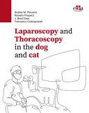
Poté, co byla v 80. letech minulého století v humánní medicíně zavedena miniinvazivní chirurgie, začaly se laparoskopické a torakoskopické zákroky prosazovat i ve veterinární medicíně. Díky menší invazivitě a rychlé pooperační rekonvalescenci se tato metoda rozšířila natolik, že se dnes stala preferovaným postupem u mnoha chirurgických zákroků.
Publikace představuje komplexní zdroj informací o nejdůležitějších miniinvazivních chirurgických postupech. Popis jednotlivých kroků operace doprovází bohatá obrazová dokumentace. Publikace je rozdělena do tří sekcí.
První sekce je věnována historii miniinvazivních chirurgických zákroků, vybavení pro tyto zákroky, zásady dodržování sepse na operačním sálu, ergonomii pracoviště, dovednosti při manipulaci v portech a fíglech při šití v průběhu laparoskopických a torakoskopických zákrocích, možnostech procvičování zručností na různých modelech. Zajímavá je kapitola o použití indocyanové zeleně k lepší vizualizaci cév v operované oblasti.
Druhá sekce se zabývá laparoskopickými operacemi. První kapitola řeší problematiku volby optimální anestezie. Za touto kapitolou je zdokumentován klinický případ řešení hypotenze, bradykardie a pneumoperitonea u feny křížence. V následující kapitole jsou podrobně popsány možnosti polohování a přípravy pacientů. V další kapitole jsou uvedeny možné komplikace při laparoskopii a jejich řešení. Detailně jsou rozpracovány metodiky odběru vzorků v dutině břišní a pánevní při laparoskopii. Každá kapitola popisující miniinvazivní zákroky je zakončena záznamem klinického případu. Tento záznam je členěn na popis případu, klíčovými body, popisem umístění operačních portů, různými triky a tipy, doporučenou pooperační péčí.Vše je bohatě ilustrováno krásnými intraoperativníi snímky vysoké kvality s popisem chirurgického postupu. Detailně jsou popsány nejčastější zákroky prováděné v dutině břišní (laparoskopická ovariohysterektomi) a pánevní (cystoskopie).
Třetí sekce zahrnuje poznatky získané při thorakoskopických vyšetřeních a zákrocích. Tato sekce je koncipována podobně jako předcházející sekce. Od indikace, selekce pacientů, předoperační vyšetření, stabilizace, příprava a polohování pacienta, zásady optimální anestezie a analgezie, možnosti vizualizace operačního pole různými zobrazovacími metodami, zvládnutí řízené ventilace, její monitoring a komplikace. Všechny tyto kroky jsou dále detailně zpracovány v následných kapitolách. Každá kapitola pomocí nákresů a fotografií znázorňuje rozmístění chirurgického týmu na operačním sále i umístění vstupních portů pro provedení laparoskopických či torakoskopických zákroků. Publikaci obohacují také osobní zkušenosti mezinárodního autorského kolektivu, který sdílí postupy dosud v odborné literatuře nepopsané, což z této knihy činí nepostradatelnou praktickou příručku jak pro běžné veterinární lékaře, tak pro zkušené chirurgy.
Na konci každé sekce knihy a vybraných kapitol je k dispozici QR kód s odkazem na seznam doporučené literatury. Pro lepší pochopení látky kniha obsahuje videa popisovaných zákroků, která jsou přístupná prostřednictvím QR kódů na webové stránce: vetminimallyinvasivesurgery.edrapublishing.com. Snad proto se v této knize hemží velkým množstvím QR kódů. Velké množství barevných fotografií, excelentní přehledná grafická úprava z této publikace vytvořilo jedinečný zdroj informací tohoto oboru.
Autor: Andrea M. Pievaroli, Roberto Properzi, J. Brad Case, Francesco Collivignarelli
| Nakladatel | Edra Publishing |
|---|---|
| ISBN | 9781957260778 |
| Vydání | 2023 |
| Vazba | pevná |
| Počet stran | 696 |
After the introduction in the 1980s of minimally invasive surgery in human medicine, laparoscopic and thoracoscopic surgery started to take hold also in veterinary medicine. Thanks to its reduced invasiveness and its fast post-operative recovery, minimally invasive surgery started to expand so quickly that nowadays it has become the preferred technique for many surgical procedures. Laparoscopy and Thoracoscopy in the dog and cat is a comprehensive book that describe all the minimally invasive surgical procedures. The description of the various surgical steps is accompanied by a wide iconography, each chapter in fact illustrates with drawings and pictures the arrangement of the surgeons in the operating room and the positioning of portals to perform laparoscopic or thoracoscopic procedures. The book is also enriched by personal experiences of international authors who shared procedures still not described in the literature making the volume a must have practical book for practitioners aswell as experienced surgeons. For a better understanding the book includes videos of the described procedures available through QR codes on vetminimallyinvasivesurgery.edrapublishing.com.
Andrea M. Pievaroli
Andrea M. Pievaroli graduated in 1992 from Università degli Studi in Milan.Since 1993 he has worked in Rome in a private referral clinic, mainly dealing with soft tissue surgery, minimally invasive surgery and orthopedics. He spent periods at the College of Veterinary Medicine of Michigan State University (USA), the College of Veterinary Medicine of Texas A&M University (USA), and the Animal Medical Center (NY, USA). Since 2000 he is particularly experienced in minimally invasive endoscopic surgery (Laparoscopy and Thoracoscopy), cystoscopy and other endoscopic techniques. He is an instructor for minimally invasive endoscopic courses and has presented at national and international congresses and events. He is a member and liaison of VES (Veterinary Endoscopy Society) and secretary of SICMIV (Italian Society of Veterinary Mini-Invasive Surgery).
Roberto Properzi
Roberto Properzi is the author of several scientific articles, mainly in the surgical field.
He has presented numerous dissertations at specialized international conferences organized by VES, SICV, SIOVET, SICEV, SLEV, ASAEE and EVENDOC. He participated in four courses in Caceres (Spain) at the CCMIJU (Jesus Uson Minimally Invasive Surgery Center), in June 2009 and January 2011 as a student, in 2018 and 2022 as an instructor. He was a lecturer at the Master of Surgery in the years 2021-2022, at the Faculty of Veterinary Medicine of Teramo. He mainly practices orthopedic and minimally invasive surgery and is particularly keen on laparoscopy, thoracoscopy and arthroscopy. He is an associate member of the following organizations: VES, ASAEE, EVENDOC, MINIMAL, SIOVET, SICV, SICMIV, SITOV, ESVOT, SLEV and SICEV. His hobbies are both the mountain, where he practices ski mountaineering and the sea, which sets sail with his motorboat. He loves beautiful company too.
J. Brad Case
J. Brad Case is a Professor of Small Animal, Minimally Invasive and Soft Tissue Surgery at the University of Florida. He obtained his DVM degree from The University of California, Davis. He did an internship in medicine and surgery at Texas A&M University. He then went on to complete a surgery residency and Master’s degree at Colorado State University. He has been a surgeon at the University of Florida since 2011. During this time, he has trained multiple surgery residents and minimally invasive surgery fellows. He has developed a vascular interventional service after training with University of Florida interventional radiologists. His translational research has been funded by the NIH and the Society of Thoracic surgeons which has led to the development of devices and procedures intended for use in humans with cardiovascular disease.
Francesco Collivignarelli
Francesco Collivignarelli graduated at the University of Perugia and started a Master’ s degree in Oncology and Oncologic Surgery at the University of Pisa and and a PhD from the University of Teramo. He did an Internship in Soft Tissue Surgery at University of Bristol Prof. Peter Holt (ECVS). After that he gained a fellowship in Oncologic Surgery at University of Torino under the supervision of Prof. Paolo Buracco (ECVS). He is the coauthor and collaborator of the book Advances in Small Animal Care vol.1 (Elsevier 2020). And also the author and coauthor of many scientific studies and scientific papers. He is often invited as guest speaker at national and international meetings. Since 2020° is the contract Professor at Master of Small Animal Surgery University of Teramo. He i s also a Lecturer at the Master of Diagnostic Image University of Teramo (2020 to date).Currenly is working as surgeon at Teaching Hospital at University of Teramo, and at AniCura at Policlinico Roma Sud. His field of interest are sentinel lymph node, minimally invasive surgery, oncologic surgery, cardiovascular surgery, oromaxillofacial oncologic surgery. He is the secretary of AIVPA, SICEV Italian associations for small animal. Professional CCR Diver
Section I. GENERAL PRINCIPLES
1. HISTORY OF VETERINARY MINIMALLY INVASIVE SURGERY
Lynetta J. Freeman
Beagle ovariohysterectomy model
Open approach to the abdomen
Trocar and clip applier testing
Expansion of preclinical research and development
Early knowledge dissemination
Early clinical application
Veterinary Endoscopy Society
Advances in education
Natural orifice transluminal endoscopic surgery (NOTES)
Single-incision laparoscopic surgery
Future of minimally invasive surgery in animals
2. LOW-IMPACT SURGERY
Luca Formaggini
3. EQUIPMENT FOR MINIMALLY INVASIVE SURGERY
Fausto Brandão, Christopher J. Chamness
Imaging systems
Light sources
Light transmitting cables
Camera control units and camera heads
Insufflators
Documentation units
Surgical monitors
Instrumentation
Electrosurgery units and vessel-sealing technology
Accessory devices
Surgical tables and racks
Other accessory units and devices for laparoscopy and thoracoscopy
4. OPERATING ROOM AND HYGIENE MEASURES
Roberto Properzi, Francesco Collivignarelli
Considerations for a minimally invasive operating room
Basic ergonomic considerations
Operating room checklist
Preparation for open surgery
Hybrid rooms
Hygiene measures
Cleaning of hands
5. ERGONOMICS IN LAPAROSCOPY AND THORACOSCOPY
Andrea M. Pievaroli
Domains of ergonomics
Operating room setup
Factors linked to musculoskeletal disorders
6. PRINCIPLES OF TRIANGULATION IN LAPAROSCOPY AND THORACOSCOPY
Andrea M. Pievaroli
Mechanical constraints
Visual restrictions
Optimal position of the rigid endoscope (T1)
Optimal position of operative instruments (T2, T3)
Relationship between the endoscope and the working instruments
7. SINGLE-INCISION (SINGLE-PORT) LAPAROSCOPIC TECHNIQUE
Jeffrey J. Runge, Joshua G. Becker
Principles of single-port surgery
Instrumentation
Optics
Single-port access methods
Equipment-specific access methods
A. Insertion techniques for specifically manufactured single‐port devices
B. Insertion technique of standard trocars for single‐port entry
C. Innovative adaptations of existing equipment
8. LAPAROSCOPIC INTRACORPOREAL AND EXTRACORPOREAL SUTURING
M. A. Sánchez Hurtado, J. Gutiérrez del Sol, F. J. Pérez Duarte
Ergonomics
Instruments and equipment
Training in laparoscopic suturing
Suture materials
Types and patterns of laparoscopic suturing
Laparoscopic intracorporeal suturing
Laparoscopic extracorporeal suturing
Additional suturing and knot tying techniques
Robotic, articulated, and mechanical devices
How many turns and throws do I need?
Knot security in laparoscopic surgery
What is the best knotting configuration?
9. LEARNING AND ADVANCING MINIMALLY INVASIVE SURGERY SKILLS
Boel A. Fransson
Basic psychomotor skills
Laparoscopic entry and basic procedure practice
Performing basic procedures in live animals
Advanced procedure training
10. INDOCYANINE GREEN INITIAL APPLICATIONS OF FLUORESCENCE-GUIDED SURGERY
Fausto Brandão, Constantin Vasile Ifteme
Fundamentals of NIR/ICG
Imaging technology
Procedure principles
Clinical applications
Case examples
Section II. LAPAROSCOPY
11. ANESTHESIA IN LAPAROSCOPIC SURGERY
Alessandra Malavasi
Anesthetic management
Postoperative management
Complications
Clinical cases
11.1 Hypotension and bradycardia at pneumoperitoneum induction in a female dog
Samuele Venuta
12. PATIENT POSITIONING, ACCESS, PORT PLACEMENT, AND PNEUMOPERITONEUM IN LAPAROSCOPY
Roberto Properzi
Patient positioning
Laparoscopic access and port placement
Pneumoperitoneum
13. COMPLICATIONS IN LAPAROSCOPY
Francesco Collivignarelli
Anatomical knowledge
Closed entry with the Veress needle technique
Open entry techniques
Laparoscopic access complications
Intraoperative complications and conversion to open techniques
Postoperative complications
14. EXPLORATORY AND DIAGNOSTIC LAPAROSCOPY
Christopher B. Thomson, Ameet Singh
Patient preparation and positioning
Operating room layout
Port placement
A. Abdominal exploration
B. Liver and gallbladder evaluation and biopsy
C. Kidney and adrenal gland evaluation and biopsy
D. Pancreas evaluation and biopsy
E. Spleen evaluation and biopsy
F. Lymph node evaluation and biopsy
G. Gastrointestinal tract evaluation and biopsy
Postoperative management
Clinical cases
14.1 Cisterna chyli ablation
David García Rubio
14.2 Laparoscopic intraperitoneal foreign
body retrieval (two-port technique)
Erin A. Gibson, William T. N. Culp
14.3 Laparoscopic exploration in a cat
Antoine Adam
14.4 Laparoscopic treatment of hemoabdomen
Michael Brückner
14.5 Minimally invasive abdominal approach to subcostal foreign body removal
Matteo Balestrieri, Boris Dalpozzo
14.6 Laparoscopic repositioning of a ventriculoperitoneal shunt catheter
Michael Brückner
14.7 Laparoscopic liver biopsy
Enrico Sponza, Francesca Molinari
15. LAPAROSCOPIC OVARIECTOMY
Andrea M. Pievaroli
Described surgical procedures
Patient preparation and positioning
Operating room layout
A. Two-port ovariectomy
B. Three-port ovariectomy
C. Single-port laparoscopic ovariectomy
Jeffrey J. Runge, Joshua G. Becker
Postoperative management
Clinical cases
15.1 Ovariectomy by transvaginal total NOTES.
Maurício Veloso Brun, Marco Augusto Machado-Silva
15.2 Two-port laparoscopic ovariectomy in the cat
Antoine Adam
15.3 Laparoscopic ovariectomy in a dog with type III sclerosing encapsulating peritonitis
Michael Brückner
15.4 Laparoscopic ovariectomy in large- and giant-breed dogs
Paolo Boschi, Roberto Properzi
15.5 Two-port laparoscopic ovariectomy without ovary suspension suture in a female dog
Giovanni Allevi
15.6 Laparoscopic ovariectomy in an African lioness (Panthera leo) using a single-port
multiple-access device
Mathieu Manassero
15.7 LESS ovariectomy using a simple
handmade access device
Marco Augusto Machado Silva, Maurício Veloso Brun
16. LAPAROSCOPY FOR OVARIAN REMNANT SYNDROME
J. Brad Case
Described surgical procedures
Patient preparation and positioning
Operating room layout
Port placement
Procedure
Postoperative management
Clinical cases
16.1 Ovarian remnant syndrome after laparoscopic ovariectomy in a cat
Paolo Boschi, Roberto Properzi
16.2 Ovarian remnant syndrome in a dog
Ivan Jevtic
17. LAPAROSCOPIC OVARIOHYSTERECTOMY
Andrea M. Pievaroli
Described surgical procedures
Patient preparation and positioning
Operating room layout
A. Two-port ovariohysterectomy
B. Three-port ovariohysterectomy
Laparoscopic ovariohysterectomy for ovarian or uterine pathologies
Postoperative management
18. LAPAROSCOPIC CRYPTORCHIDECTOMY
Andrea M. Pievaroli
Patient preparation and positioning
Operating room layout
A. Two-port laparoscopic cryptorchidectomy
B. Single-port laparoscopic cryptorchidectomy
Jeffrey J. Runge, Joshua G. Becker
Postoperative management
Clinical cases
18.1 Testicular torsion in a cryptorchid dog
Andrea M. Pievaroli
18.2 Laparoscopic cryptorchidectomy
D. L. Casas García
19. LAPAROSCOPIC-ASSISTED CYSTOSCOPY AND PERCUTANEOUS SUPRAPUBIC CYSTOSCOPY
Andrea M. Pievaroli
Patient preparation and positioning
Operating room layout
A. Laparoscopic-assisted cystoscopy
B. Percutaneous suprapubic cystoscopy or percutaneous cystolithotomy (PCCL)
C. Single-port percutaneous cystolithotomy
Jeffrey J. Runge, Joshua G. Becker
Postoperative management
Clinical cases
19.1 Percutaneous cystoscopic cystolithotomy in a Yorkshire Terrier
Christopher B. Thomson
19.2 Laparoscopic-assisted cystostomy in a Chihuahua
D. L. Casas García
20. LAPAROSCOPIC CYSTOTOMY
Andrea M. Pievaroli
Patient preparation and positioning
Operating room layout
Port placement
Procedure
Postoperative management
21. LAPAROSCOPIC CYSTOPEXY
J. Brad Case
Described surgical procedures
Patient preparation and positioning
Operating room layout
Port placement
Procedure
Postoperative management
22. LAPAROSCOPIC-ASSISTED VASOPEXY
Massimo Barilli
Patient preparation and positioning
Three-port laparoscopic-assisted vasopexy
Postoperative management
Clinical cases
22.1 Laparoscopic reversible neutering in a dog
Roberto Giaconella, Emanuela Vitali, Veronica Giaconella
23. LAPAROSCOPIC URETERONEPHRECTOMY
Roberto Properzi
Described surgical procedures
Patient preparation and positioning
Operating room layout
Port placement
Procedure
Postoperative management
Clinical cases
23.1 Laparoscopic omentalization of a renal cyst
Michael Brückner
23.2 Laparoscopic perinephric pseudocyst resection
Justin B. Ganjei
23.3 Laparoscopic renal cyst deroofing, fulguration, and omentalization
Mathieu Manassero, Bastien Dekerle
24. CURRENT CONCEPTS IN MINIMALLY INVASIVE NEPHROLITHOTOMY
Fausto Brandão
Described surgical procedures
Patient selection
Equipment
Operating room layout
Modified surgical-assisted nephrolithotomy technique
Postoperative management
Clinical cases
24.1 Nephron-sparing nephrotomy to remove large uroliths in dogs
Maurício Veloso Brun
24.2 Nephron-sparing nephrotomy for the treatment of Dioctophyme renale parasitosis
Maurício Veloso Brun
25. LAPAROSCOPIC APPROACH TO URETEROLITHIASIS
J. Gutiérrez del Sol, F. J. Pérez Duarte, M. A. Sánchez Hurtado
Patient preparation and positioning
Operating room layout
Port placement
Procedure
Postoperative management
Clinical cases
25.1 Ureterotomy for stone removal
David García Rubio
26. LAPAROSCOPIC NEOURETEROCYSTOSTOMY
Felipe Lillo
Described surgical procedures
Patient preparation and positioning
Operating room layout
Port placement
Procedure
Postoperative management
Clinical cases
26.1 Laparoscopy-assisted cutaneous ureterostomy (LACU) in a canine patient with prostatic carcinoma
Giovanni Allevi
27. LAPAROSCOPIC ARTIFICIAL URETHRAL SPHINCTER (AUS) IMPLANTATION
F. J. Pérez Duarte, Maurici Batalla, M. A. Sánchez Hurtado, J. Gutiérrez del Sol
Patient preparation and positioning
Operating room layout
Port placement
Procedure
Postoperative management
28. LAPAROSCOPIC PROSTATECTOMY
Felipe Lillo, Francesco Collivignarelli
Described surgical procedures
Patient preparation and positioning
Operating room layout
Port placement
Procedure
Postoperative management
29. LAPAROSCOPIC OMENTALIZATION OF PARAPROSTATIC CYSTS
Andrea M. Pievaroli
Patient preparation and positioning
Operating room layout
Port placement
Procedure
Postoperative management
Clinical cases
29.1 Omentalization of a prostatic abscess
David García Rubio
29.2 Omentalization of a paraprostatic cyst in a dog
Massimo Barilli
30. LAPAROSCOPIC GASTROPEXY AND LAPAROSCOPIC-ASSISTED GASTROPEXY
J. Brad Case, Roberto Properzi
Described surgical procedures
A. Intracorporeal laparoscopic gastropexy I
J. Brad Case
B. Intracorporeal laparoscopic gastropexy II
Roberto Properzi
C. Laparoscopic-assisted gastropexy
D. Single-port laparoscopic-assisted incisional gastropexy
Jeffrey J. Runge, Joshua G. Becker
Postoperative management
Clinical cases
30.1 Total laparoscopic sutureless gastropexy
with absorbable straps in dogs
Luca Lacitignola
30.2 Intracorporeally sutured laparoscopic gastropexy using an abdominal wall incision made at right angles to the rotation direction of the suturing needle
Motoki Kondo
30.3 Laparoscopic gastropexy in a dog
Antoine Adam
30.4 Total laparoscopic gastropexy (two-port technique using Endostitch)
Justin B. Ganjei
30.5 Treatment of lower esophageal sphincter incompetence in a cat using total laparoscopic gastropexy
Roberto Giaconella, Veronica Giaconella
31. LAPAROSCOPIC-ASSISTED GASTROTOMY
Andrea M. Pievaroli
Patient preparation and positioning
A. Two-port laparoscopic-assisted gastrotomy
B. Single-port laparoscopic-assisted gastrotomy
Jeffrey J. Runge, Joshua G. Becker
Postoperative management
32. SINGLE-PORT LAPAROSCOPIC-ASSISTED INTESTINAL SURGERY
Jeffrey J. Runge, Joshua G. Becker
Patient preparation and positioning
Operating room layout
Port placement
Procedure
Postoperative management
33. LAPAROSCOPIC-ASSISTED COLOPEXY
Massimo Barilli
Patient preparation and positioning
Operating room layout
A. Two-port laparoscopic-assisted colopexy
B. Three-port laparoscopic-assisted colopexy and vasopexy
Postoperative management
Clinical cases
33.1 Totally laparoscopic colopexy in a cat
Andrea M. Pievaroli
33.2 Laparoscopic colopexy and ovariectomy in a dog
Christopher B. Thomson
34. LAPAROSCOPIC LIVER SURGERY
David García Rubio, Edoardo Poggi, Luciano Borghetti
Patient preparation and positioning
Operating room layout
A. Right medial hepatectomy in a cat using a bipolar sealing device
B. Laparoscopic partial left lateral hepatectomy
C. Laparoscopic partial left medial hepatectomy
Postoperative management
Clinical cases
34.1 Laparoscopic excisional liver biopsy in a dog
Przemysław Prza˛dka, Bartłomiej Liszka, Joanna Tunikowska, Stanisław Dzimira, Dariusz Patkowski
34.2 Laparoscopic excision of a hepatic cyst
and omentopexy in a Persian cat
Giovanni Allevi
35. LAPAROSCOPIC MICROWAVE ABLATION OF THE LIVER
J. Brad Case, Beau B. Toskich
Described surgical procedures
Patient preparation and positioning
Operating room layout
Port placement
Procedure
Postoperative management
36. LAPAROSCOPIC CHOLECYSTECTOMY
Eric Monnet
Patient preparation and positioning
Operating room layout
Port placement
Procedure
A. Dissection from the cystic duct
B. Dissection from the apex of the gallbladder
C. Intraoperative cholangiogram
Postoperative management
Clinical cases
36.1 Single-port laparoscopic cholecystectomy technique in dogs
Jeffrey J. Runge, Joshua G. Becker
36.2 Laparoscopic cholecystectomy and intraoperative bile duct flushing for extrahepatic biliary obstruction in a dog
Hiroo Kanai
36.3 Cholecystectomy and bile duct flushing
Ikuya Ehara
36.4 Laparoscopic cholecystectomy in a dog using the subserosal layer dissection technique
Motoki Kondo
36.5 Cholecystectomy
David García Rubio
36.6 Cholecystectomy by three-port access and “cold” dissection
Maurício Veloso Brun
36.7 Cholecystectomy in a dog
F. J. Pérez Duarte, J. Gutiérrez del Sol, M. A. Sánchez Hurtado
36.8 Laparoscopic cholecystectomy in a dog
Ivan Jevtic
36.9 Fundus-first cholecystectomy
Christopher B. Thomson
37. LAPAROSCOPIC TREATMENT OF EXTRAHEPATIC PORTOSYSTEMIC SHUNT
Francesco Collivignarelli, Roberto Properzi, Maurício Veloso Brun, Robson Pasquale
A. Laparoscopic treatment of extrahepatic portosystemic shunt with cellophane band
Francesco Collivignarelli, Roberto Properzi
B. Laparoscopic treatment of extrahepatic portosystemic shunt with ameroid constrictor
Maurício Veloso Brun, Robson Pasquale
Clinical cases
37.1 Splenocaval shunt
David García Rubio
37.2 Laparoscopic attenuation of a splenophrenic shunt in a dog
Edoardo Poggi, Luciano Borghetti, Francesca Izzo
37.3 Laparoscopic attenuation of a splenocaval shunt in a dog
Edoardo Poggi, Francesca Izzo, Luciano Borghetti
37.4 Laparoscopic attenuation of a left intrahepatic portosystemic shunt in a cat
Edoardo Poggi, Filippo Cinti, Francesca Izzo
37.5 Laparoscopic portosystemic shunt attenuation in a dog
F. J. Pérez Duarte, J. Gutiérrez del Sol, M. A. Sánchez Hurtado
38. LAPAROSCOPIC PANCREATIC SURGERY
S. A. van Nimwegen, F. O. Buishand, J. Kirpensteijn
Described surgical procedures
A. Three-port pancreatic biopsy
B. Three-port excision of pancreatic lesions, excluding lesions in the distal left pancreatic lobe
C. Three-port excision of pancreatic lesions in the distal left pancreatic lobe
D. Laparoscopic approach for insulinoma going beyond the pancreas
Postoperative management
Clinical cases
38.1 Pancreatectomy in a case of insulinoma
David García Rubio
38.2 Laparoscopic right lateral pancreatic lobectomy in a dog
Edoardo Poggi, Filippo Cinti, Francesca Izzo
38.3 Laparoscopic omentalization of a pancreatic cyst in a cat
Michael Brückner
39. LAPAROSCOPIC SPLENECTOMY
Maurici Batalla
Patient preparation and positioning
Operating room layout
Port placement
A. Multiport laparoscopic splenectomy
B. Single-incision laparoscopic splenectomy
C. Laparoscopic-assisted splenectomy
D. Hand-assisted splenectomy
Postoperative management
Clinical cases
39.1 Laparoscopic splenectomy in a small dog
Andrea M. Pievaroli
39.2 Splenectomy
D. L. Casas García
39.3 Laparoscopic splenectomy for hemangiosarcoma
Fabien Collard
40. LAPAROSCOPIC ADRENALECTOMY
Francesco Collivignarelli, Felipe Lillo
Endocrine testing
Diagnostic imaging
Described surgical procedures
A. Adrenalectomy in sternal recumbency
B. Adrenalectomy in lateral recumbency
Postoperative management
C. Retroperitoneal adrenalectomy
Clinical cases
40.1 Right laparoscopic adrenalectomy
William T. N. Culp
40.2 Laparoscopic right adrenalectomy for adrenal adenocarcinoma in a dog
Hiroo Kanai
40.3 Right adrenalectomy in a Yorkshire Terrier dog
David García Rubio
40.4 Left adrenalectomy in a dog
Ikuya Ehara
40.5 Laparoscopic right adrenalectomy
Przemysław Przadka, Stanisław Dzimira, Agnieszka Antończyk, Łukasz Juźwiak, Bartłomiej Liszka
41. LAPAROSCOPIC DIAPHRAGMATIC HERNIORRHAPHY
J. Brad Case, Maurício Veloso Brun
Described surgical procedures
A. Three-port laparoscopic diaphragmatic herniorrhaphy
B. Three-port gasless laparoscopic diaphragmatic herniorrhaphy
Postoperative management
Clinical cases
41.1 Diaphragmatic hernia in a cat
David García Rubio
42. LAPAROSCOPIC HIATAL HERNIORRHAPHY
J. Brad Case
Described surgical procedures
Patient preparation and positioning
Operating room layout
Port placement
Procedure
Postoperative management
43. LAPAROSCOPIC INGUINAL HERNIORRHAPHY
Alexander Chernov
Described surgical procedures
Patient preparation and positioning
Operating room layout
Port placement
Procedure
Postoperative management
Clinical cases
43.1 Laparoscopic-assisted percutaneous herniorrhaphy in dogs using the PIRS technique and laparoscopic ovariectomy
Przemysław Przadka, Bartłomiej Liszka, Agnieszka Antończyk, Zdzisław Kiełbowicz, Dariusz Patkowski
43.2 Laparoscopic-assisted percutaneous
inguinal herniorrhaphy
F. Martínez Gomariz, C. Sánchez, J. D. Carrillo
44. LAPAROSCOPIC PERINEAL HERNIORRHAPHY
Alexander Chernov
Patient preparation and positioning
Operating room layout
Port placement
Procedure
Postoperative management
45. LAPAROSCOPIC PERITONEOPERICARDIAL HERNIORRHAPHY
Valery F. Scharf
Patient preparation and positioning
Operating room layout
Port placement
Procedure
Postoperative management
Clinical cases
45.1 Laparoscopic treatment of peritoneal- pericardial diaphragmatic hernia in a puppy dog
Andrea M. Pievaroli
45.2 Laparoscopic repair of pericardial diaphragmatic hernia in a dog
Hiroo Kanai
Section III. THORACOSCOPY
46. ANESTHESIA IN THORACOSCOPY
Elisa Bortolami
Indications for thoracoscopic procedures
Patient selection
Preoperative work-up
Patient stabilization
Patient preparation and positioning
Anesthesia
Analgesia
Visualization of the surgical field and lung separation
Pathophysiology of one-lung ventilation
Management of ventilation
Complications during one-lung ventilation
Monitoring
3
Postoperative complications
Postoperative management
47. PATIENT POSITIONING, ACCESS, AND PORT PLACEMENT IN THORACOSCOPY
Roberto Properzi
Patient positioning
Thoracoscopic access and port placement
48. CONTRAINDICATIONS AND COMPLICATIONS IN THORACOSCOPY
J. Brad Case
Strategic avoidance of complications
Most frequent complications
Monitoring for complications
49. EXPLORATORY AND DIAGNOSTIC THORACOSCOPY
Francisco Miguel Sánchez-Margallo, Isabel López-Agudelo, Manuel Ramón González-Portillo, David Durán-Rey
A. Thoracoscopic exploration
B. Thoracic biopsy
Postoperative management
Clinical cases
49.1 Thoracoscopy in combination with digestive endoscopy for the diagnosis of intrathoracic neoformations
Francesca Molinari, Enrico Sponza, Giacomo Federici
49.2 Gunshot wound to the chest cavity in a dog
Osichkin P. V., Kuznetsov P. A.
50. THORACOSCOPIC PERICARDIECTOMY
Roberto Bussadori, Andrea Pievaroli, Roberto Properzi, Deborah De Bastiani
A. Pericardial window in dorsal recumbency
B. Pericardial window in lateral recumbency
C. Subtotal pericardiectomy
Postoperative management
Clinical cases
50.1 Two-port thoracoscopic pericardiectomy as a novel technique in flat-chested dogs with reduced thoracic working spaces
Andrea M. Pievaroli
50.2 Thoracoscopic subphrenic (subtotal) pericardiectomy
Giovanni Allevi
51. THORACOSCOPIC THORACIC DUCT LIGATION FOR TREATMENT OF CHYLOTHORAX
J. Brad Case
Described surgical procedures
Patient preparation and positioning
Operating room layout
Port placement
Procedure
Postoperative management
Clinical cases
51.1 Bilateral en bloc thoracic duct ligation in combination with pericardiectomy by video-assisted thoracoscopic surgery for idiopathic chylothorax in a dog
Hiroo Kanai
51.2 Thoracoscopic thoracic duct ligation for idiopathic chylothorax
F. J. Pérez Duarte, J. Gutiérrez del Sol, M. A. Sánchez Hurtado
51.3 Thoracic duct ligation and pericardiectomy in a cat with chylothorax
Antoine Adam
51.4 Thoracoscopic thoracic duct ligation and methylene blue coloration of mesenteric lymph nodes to confirm occlusion
Leonardo Cavaliere, Flavia Catanzaro
51.5 Endoscopic-assisted surgery for idiopathic chylothorax in a domestic shorthair cat
Yi Fu Chang, Ming Ju Huang
51.6 Chylothorax associated with a peritoneopericardial diaphragmatic hernia in a dog
Andrea M. Pievaroli
52. THORACOSCOPIC APPROACH TO THE MANAGEMENT OF PYOTHORAX
Eric Monnet
Operating room layout
Port placement
Procedure
Postoperative management
53. THORACOSCOPIC LUNG LOBECTOMY
Philipp D. Mayhew, Andrea Pievaroli, Roberto Properzi, Francesco Collivignarelli
Patient preparation and positioning
Operating room layout
A. Thoracoscopic lung lobectomy
B. Thoracoscopically assisted lung lobectomy
Postoperative management
Clinical cases
53.1 Hilar lung lobectomy
Ikuya Ehara
53.2 Video-assisted thoracoscopic surgery pulmonary lobectomy
David García Rubio
53.3 Thoracoscopic lung lobectomy for lung lobe torsion
Roberto Bussadori, Deborah De Bastiani
53.4 Thoracoscopic lobectomy of the right middle lung lobe in a dog
Li-Ren Wang, Jung-Hsien Sun,Yi-Fu Chang
54. THORACOSCOPIC APPROACH TO VASCULAR RING ANOMALIES
J. Brad Case
Described surgical procedures
Patient preparation and positioning
Operating room layout
Port placement
Procedure
Postoperative management
Clinical cases
54.1 Thoracoscopic approach combined with esophagoscopy for the surgical correction of a persistent right aortic arch
F. J. Pérez Duarte, J. Gutiérrez del Sol, M. A. Sánchez Hurtado
55. THORACOSCOPIC APPROACH TO MEDIASTINAL MASS EXCISION
J. Brad Case
Patient preparation and positioning
Operating room layout
Port placement
Procedure
Postoperative management
Clinical cases
55.1 Thoracoscopic mediastinal lymph node excision
Justin B. Ganjei
55.2 Thoracoscopic treatment in a cat with thymoma-associated exfoliative dermatitis
Francesca Izzo, Luciano Borghetti
55.3 Thoracoscopic resection of a cranial mediastinal mass in a dog
Li-Ren Wang, Jung-Hsien Sun
55.4 Thoracoscopic excision of a mediastinal ectopic thyroid carcinoma
Boris Dalpozzo
56. THORACOSCOPIC AURICULECTOMY
J. Brad Case
Described surgical procedures
Patient preparation and positioning
Operating room layout
Port placement
Procedure
Postoperative management
Clinical cases
56.1 Thoracoscopic right auricular tumor excision using a four-port technique
Justin B. Ganjei
56.2 Surgical variation on auricular mass removal technique
Boris Dalpozzo
57. THORACOSCOPIC FOREIGN BODY REMOVAL
Boris Dalpozzo
Diagnosis
Preoperative considerations
Patient preparation and positioning
Operating room layout
Port placement
Procedure
Postoperative management
58. THORACOSCOPIC PLACEMENT OF EPICARDIAL PACEMAKERS
Lucie Bonneau, Stéphane Libermann
Patient preparation and positioning
Operating room layout
Port placement
Procedure
Postoperative management
59. THORACOSCOPIC-ASSISTED PLEURAL PORT PLACEMENT
Francesco Collivignarelli, Kumiko Ishigaki
A. Pleural port placement following pericardiectomy
B. Pleural port placement for the management of malignant thoracic effusion
Postoperative management
Clinical cases
59.1 Pleural port implantation for the management of malignant pleural effusion due to metastatic mammary carcinoma in a dog
Giulia Dolce, Flavia Catanzaro
| Section I. GENERAL PRINCIPLES | 1 |
| 1. HISTORY OF VETERINARY MINIMALLY INVASIVE SURGERY | 3 |
| Beagle ovariohysterectomy model | 4 |
| Open approach to the abdomen | 4 |
| Trocar and clip applier testing | 4 |
| Expansion of preclinical research and development | 5 |
| Early knowledge dissemination | 6 |
| Early clinical application | 7 |
| Veterinary Endoscopy Society | 7 |
| Advances in education | 7 |
| Natural orifice transluminal endoscopic surgery (NOTES) | 8 |
| Single-incision laparoscopic surgery | 8 |
| Future of minimally invasive surgery in animals | 8 |
| 2. LOW-IMPACT SURGERY | 9 |
| 3. EQUIPMENT FOR MINIMALLY INVASIVE SURGERY | 12 |
| Imaging systems | 12 |
| Light sources | 12 |
| Light transmitting cables | 13 |
| Camera control units and camera heads | 13 |
| Insufflators | 14 |
| Documentation units | 14 |
| Surgical monitors | 14 |
| Instrumentation | 15 |
| Electrosurgery units and vessel-sealing technology | 20 |
| Accessory devices | 20 |
| Surgical tables and racks | 20 |
| Other accessory units and devices for laparoscopy and thoracoscopy | 21 |
| 4. OPERATING ROOM AND HYGIENE MEASURES | 27 |
| Considerations for a minimally invasive operating room | 27 |
| Basic ergonomic considerations | 27 |
| Operating room checklist | 27 |
| Preparation for open surgery | 28 |
| Hybrid rooms | 28 |
| Hygiene measures | 28 |
| Cleaning of hands | 28 |
| 5. ERGONOMICS IN LAPAROSCOPY AND THORACOSCOPY | 30 |
| Domains of ergonomics | 31 |
| Operating room setup | 31 |
| Factors linked to musculoskeletal disorders | 31 |
| 6. PRINCIPLES OF TRIANGULATION IN LAPAROSCOPY AND THORACOSCOPY | 37 |
| Mechanical constraints | 38 |
| Visual restrictions | 39 |
| Optimal position of the rigid endoscope (T1) | 40 |
| Optimal position of operative instruments (T2, T3) | 41 |
| Relationship between the endoscope and the working instruments | 43 |
| 7. SINGLE-INCISION (SINGLE-PORT) LAPAROSCOPIC TECHNIQUE | 45 |
| Principles of single-port surgery | 46 |
| Instrumentation | 46 |
| Optics | 46 |
| Single-port access methods | 47 |
| Equipment-specific access methods | 47 |
| A. Insertion techniques for specifically manufactured single‐port devices | 48 |
| B. Insertion technique of standard trocars for single‐port entry | 51 |
| C. Innovative adaptations of existing equipment | 52 |
| 8. LAPAROSCOPIC INTRACORPOREAL AND EXTRACORPOREAL SUTURING | 53 |
| Ergonomics | 54 |
| Instruments and equipment | 54 |
| Training in laparoscopic suturing | 58 |
| Suture materials | 59 |
| Types and patterns of laparoscopic suturing | 59 |
| Laparoscopic intracorporeal suturing | 60 |
| Laparoscopic extracorporeal suturing | 77 |
| Additional suturing and knot tying techniques | 88 |
| Robotic, articulated, and mechanical devices | 89 |
| How many turns and throws do I need? Knot security in laparoscopic surgery | 90 |
| What is the best knotting configuration? | 90 |
| 9. LEARNING AND ADVANCING MINIMALLY INVASIVE SURGERY SKILLS | 92 |
| Basic psychomotor skills | 92 |
| Laparoscopic entry and basic procedure practice | 93 |
| Performing basic procedures in live animals | 95 |
| Advanced procedure training | 95 |
| 10. INDOCYANINE GREEN INITIAL APPLICATIONS OF FLUORESCENCE-GUIDED SURGERY | 96 |
| Fundamentals of NIR/ICG | 96 |
| Imaging technology | 97 |
| Procedure principles | 99 |
| Clinical applications | 100 |
| Case examples | 100 |
| Section II. LAPAROSCOPY | 109 |
| 11. ANESTHESIA IN LAPAROSCOPIC SURGERY | 111 |
| Anesthetic management | 113 |
| Postoperative management | 113 |
| Complications | 114 |
| Clinical cases 11.1 Hypotension and bradycardia at pneumoperitoneum induction in a female dog | 116 |
| 12. PATIENT POSITIONING, ACCESS, PORT PLACEMENT, AND PNEUMOPERITONEUM IN LAPAROSCOPY | 117 |
| Patient positioning | 117 |
| Laparoscopic access and port placement | 117 |
| Pneumoperitoneum | 120 |
| 13. COMPLICATIONS IN LAPAROSCOPY | 121 |
| Anatomical knowledge | 121 |
| Closed entry with the Veress needle technique | 121 |
| Open entry techniques | 122 |
| Laparoscopic access complications | 123 |
| Intraoperative complications and conversion to open techniques | 123 |
| Postoperative complications | 124 |
| 14. EXPLORATORY AND DIAGNOSTIC LAPAROSCOPY | 125 |
| Patient preparation and positioning | 126 |
| Operating room layout | 126 |
| Port placement | 126 |
| A. Abdominal exploration | 128 |
| B. Liver and gallbladder evaluation and biopsy | 130 |
| C. Kidney and adrenal gland evaluation and biopsy | 131 |
| D. Pancreas evaluation and biopsy | 133 |
| E. Spleen evaluation and biopsy | 133 |
| F. Lymph node evaluation and biopsy | 134 |
| G. Gastrointestinal tract evaluation and biopsy | 135 |
| Postoperative management | 136 |
| Clinical cases 14.1 Cisterna chyli ablation | 137 |
| 14.2 Laparoscopic intraperitoneal foreign body retrieval (two-port technique) | 139 |
| 14.3 Laparoscopic exploration in a cat | 140 |
| 14.4 Laparoscopic treatment of hemoabdomen | 143 |
| 14.5 Minimally invasive abdominal approach to subcostal foreign body removal | 145 |
| 14.6 Laparoscopic repositioning of a ventriculoperitoneal shunt catheter | 147 |
| 14.7 Laparoscopic liver biopsy | 148 |
| 15. LAPAROSCOPIC OVARIECTOMY | 151 |
| Described surgical procedures | 152 |
| Patient preparation and positioning | 152 |
| Operating room layout | 152 |
| A. Two-port ovariectomy | 155 |
| B. Three-port ovariectomy | 159 |
| C. Single-port laparoscopic ovariectomy | 161 |
| Postoperative management | 163 |
| Clinical cases 15.1 Ovariectomy by transvaginal total NOTES. | 164 |
| 15.2 Two-port laparoscopic ovariectomy in the cat | 167 |
| 15.3 Laparoscopic ovariectomy in a dog with type III sclerosing encapsulating peritonitis | 169 |
| 15.4 Laparoscopic ovariectomy in large- and giant-breed dogs | 171 |
| 15.5 Two-port laparoscopic ovariectomy without ovary suspension suture in a female dog | 172 |
| 15.6 Laparoscopic ovariectomy in an African lioness (Panthera leo) using a single-port multiple-access device | 174 |
| 15.7 LESS ovariectomy using a simple handmade access device | 176 |
| 16. LAPAROSCOPY FOR OVARIAN REMNANT SYNDROME | 180 |
| Described surgical procedures | 180 |
| Patient preparation and positioning | 181 |
| Operating room layout | 181 |
| Port placement | 182 |
| Procedure | 183 |
| Postoperative management | 184 |
| Clinical cases 16.1 Ovarian remnant syndrome after laparoscopic ovariectomy in a cat | 185 |
| 16.2 Ovarian remnant syndrome in a dog | 187 |
| 17. LAPAROSCOPIC OVARIOHYSTERECTOMY | 189 |
| Described surgical procedures | 489 |
| Patient preparation and positioning | 190 |
| Operating room layout | 191 |
| A. Two-port ovariohysterectomy | 192 |
| B. Three-port ovariohysterectomy | 196 |
| Laparoscopic ovariohysterectomy for ovarian or uterine pathologies | 198 |
| Postoperative management | 199 |
| 18. LAPAROSCOPIC CRYPTORCHIDECTOMY | 203 |
| Patient preparation and positioning | 203 |
| Operating room layout | 203 |
| A. Two-port laparoscopic cryptorchidectomy | 203 |
| B. Single-port laparoscopic cryptorchidectomy | 208 |
| Postoperative management | 210 |
| Clinical cases 18.1 Testicular torsion in a cryptorchid dog | 211 |
| 18.2 Laparoscopic cryptorchidectomy | 212 |
| 19. LAPAROSCOPIC-ASSISTED CYSTOSCOPY AND PERCUTANEOUS SUPRAPUBIC CYSTOSCOPY | 215 |
| Patient preparation and positioning | 215 |
| Operating room layout | 216 |
| A. Laparoscopic-assisted cystoscopy | 216 |
| B. Percutaneous suprapubic cystoscopy or percutaneous cystolithotomy (PCCL) | 221 |
| C. Single-port percutaneous cystolithotomy | 225 |
| Postoperative management | 227 |
| Clinical cases 19.1 Percutaneous cystoscopic cystolithotomy in a Yorkshire Terrier | 228 |
| 19.2 Laparoscopic-assisted cystostomy in a Chihuahua | 230 |
| 20. LAPAROSCOPIC CYSTOTOMY | 233 |
| Patient preparation and positioning | 233 |
| Operating room layout | 233 |
| Port placement | 233 |
| Procedure | 234 |
| Postoperative management | 237 |
| 21. LAPAROSCOPIC CYSTOPEXY | 238 |
| Described surgical procedures | 238 |
| Patient preparation and positioning | 238 |
| Operating room layout | 239 |
| Port placement | 239 |
| Procedure | 239 |
| Postoperative management | 242 |
| 22. LAPAROSCOPIC-ASSISTED VASOPEXY | 243 |
| Patient preparation and positioning | 243 |
| Three-port laparoscopic-assisted vasopexy | 243 |
| Postoperative management | 245 |
| Clinical cases 22.1 Laparoscopic reversible neutering in a dog | 247 |
| 23. LAPAROSCOPIC URETERONEPHRECTOMY | 249 |
| Described surgical procedures | 249 |
| Patient preparation and positioning | 249 |
| Operating room layout | 249 |
| Port placement | 251 |
| Procedure | 251 |
| Postoperative management | 253 |
| Clinical cases 23.1 Laparoscopic omentalization of a renal cyst | 254 |
| 23.2 Laparoscopic perinephric pseudocyst resection | 256 |
| 23.3 Laparoscopic renal cyst deroofing, fulguration, and omentalization | 257 |
| 24. CURRENT CONCEPTS IN MINIMALLY INVASIVE NEPHROLITHOTOMY | 260 |
| Described surgical procedures | 261 |
| Patient selection | 262 |
| Equipment | 262 |
| Operating room layout | 263 |
| Modified surgical-assisted nephrolithotomy technique | 264 |
| Postoperative management | 267 |
| Clinical cases 24.1 Nephron-sparing nephrotomy to remove large uroliths in dogs | 268 |
| 24.2 Nephron-sparing nephrotomy for the treatment of Dioctophyme renale parasitosis | 271 |
| 25. LAPAROSCOPIC APPROACH TO URETEROLITHIASIS | 273 |
| Patient preparation and positioning | 274 |
| Operating room layout | 275 |
| Port placement | 275 |
| Procedure | 276 |
| Postoperative management | 279 |
| Clinical cases 25.1 Ureterotomy for stone removal | 280 |
| 26. LAPAROSCOPIC NEOURETEROCYSTOSTOMY | 282 |
| Described surgical procedures | 282 |
| Patient preparation and positioning | 283 |
| Operating room layout | 283 |
| Port placement | 283 |
| Procedure | 284 |
| Postoperative management | 287 |
| Clinical cases 26.1 Laparoscopy-assisted cutaneous ureterostomy (LACU) in a canine patient with prostatic carcinoma | 288 |
| 27. LAPAROSCOPIC ARTIFICIAL URETHRAL SPHINCTER (AUS) IMPLANTATION | 290 |
| Patient preparation and positioning | 291 |
| Operating room layout | 291 |
| Port placement | 292 |
| Procedure | 292 |
| Postoperative management | 297 |
| 28. LAPAROSCOPIC PROSTATECTOMY | 298 |
| Described surgical procedures | 298 |
| Patient preparation and positioning | 299 |
| Operating room layout | 299 |
| Port placement | 300 |
| Procedure | 300 |
| Postoperative management | 303 |
| 29. LAPAROSCOPIC OMENTALIZATION OF PARAPROSTATIC CYSTS | 304 |
| Patient preparation and positioning | 305 |
| Operating room layout | 305 |
| Port placement | 306 |
| Procedure | 306 |
| Postoperative management | 310 |
| Clinical cases 29.1 Omentalization of a prostatic abscess | 311 |
| 29.2 Omentalization of a paraprostatic cyst in a dog | 313 |
| 30. LAPAROSCOPIC GASTROPEXY AND LAPAROSCOPIC-ASSISTED GASTROPEXY | 315 |
| Described surgical procedures | 315 |
| A. Intracorporeal laparoscopic gastropexy I | 316 |
| B. Intracorporeal laparoscopic gastropexy II | 319 |
| C. Laparoscopic-assisted gastropexy | 322 |
| D. Single-port laparoscopic-assisted incisional gastropexy | 325 |
| Postoperative management | 326 |
| Clinical cases 30.1 Total laparoscopic sutureless gastropexy with absorbable straps in dogs | 327 |
| 30.2 Intracorporeally sutured laparoscopic gastropexy using an abdominal wall incision made at right angles to the rotation direction of the suturing needle | 328 |
| 30.3 Laparoscopic gastropexy in a dog | 331 |
| 30.4 Total laparoscopic gastropexy (two-port technique using Endostitch) | 333 |
| 30.5 Treatment of lower esophageal sphincter incompetence in a cat using total laparoscopic gastropexy | 335 |
| 31. LAPAROSCOPIC-ASSISTED GASTROTOMY | 337 |
| Patient preparation and positioning | 337 |
| A. Two-port laparoscopic-assisted gastrotomy | 338 |
| B. Single-port laparoscopic-assisted gastrotomy | 341 |
| Postoperative management | 343 |
| 32. SINGLE-PORT LAPAROSCOPIC-ASSISTED INTESTINAL SURGERY | 344 |
| Patient preparation and positioning | 344 |
| Operating room layout | 344 |
| Port placement | 345 |
| Procedure | 345 |
| Postoperative management | 347 |
| 33. LAPAROSCOPIC-ASSITED COLOPEXY | 349 |
| Patient preparation and positioning | 349 |
| Operating room layout | 349 |
| A. Two-port laparoscopic-assisted colopexy | 349 |
| B. Three-port laparoscopic-assisted colopexy and vasopexy | 352 |
| Postoperative management | 353 |
| Clinical cases 33.1 Totally laparoscopic colopexy in a cat | 354 |
| 33.2 Laparoscopic colopexy and ovariectomy in a dog | 356 |
| 34. LAPAROSCOPIC LIVER SURGERY | 358 |
| Patient preparation and positioning | 359 |
| Operating room layout | 359 |
| A. Right medial hepatectomy in a cat using a bipolar sealing device | 359 |
| B. Laparoscopic partial left lateral hepatectomy | 362 |
| C. Laparoscopic partial left medial hepatectomy | 364 |
| Postoperative management | 365 |
| Clinical cases 34.1 Laparoscopic excisional liver biopsy in a dog | 368 |
| 34.2 Laparoscopic excision of a hepatic cyst and omentopexy in a Persian cat | 370 |
| 35. LAPAROSCOPIC MICROWAVE ABLATION OF THE LIVER | 372 |
| Described surgical procedures | 372 |
| Patient preparation and positioning | 373 |
| Operating room layout | 373 |
| Port placement | 376 |
| Procedure | 376 |
| Postoperative management | 378 |
| 36. LAPAROSCOPIC CHOLECYSTECTOMY | 380 |
| Patient preparation and positioning | 380 |
| Operating room layout | 380 |
| Port placement | 380 |
| Procedure | 382 |
| A. Dissection from the cystic duct | 383 |
| B. Dissection from the apex of the gallbladder | 385 |
| C. Intraoperative cholangiogram | 385 |
| Postoperative management | 385 |
| Clinical cases 36.1 Single-port laparoscopic cholecystectomy technique in dogs | 387 |
| 36.2 Laparoscopic cholecystectomy and intraoperative bile duct flushing for extrahepatic biliary obstruction in a dog | 390 |
| 36.3 Cholecystectomy and bile duct flushing | 392 |
| 36.4 Laparoscopic cholecystectomy in a dog using the subserosal layer dissection technique | 394 |
| 36.5 Cholecystectomy | 396 |
| 36.6 Cholecystectomy by three-port access and “cold” dissection | 398 |
| 36.7 Cholecystectomy in a dog | 402 |
| 36.8 Laparoscopic cholecystectomy in a dog | 404 |
| 37. LAPAROSCOPIC TREATMENT OF EXTRAHEPATIC PORTOSYSTEMIC SHUNT | 408 |
| A. Laparoscopic treatment of extrahepatic portosystemic shunt with cellophane band | 408 |
| B. Laparoscopic treatment of extrahepatic portosystemic shunt with ameroid constrictor | 412 |
| Clinical cases 37.1 Splenocaval shunt | 418 |
| 37.2 Laparoscopic attenuation of a splenophrenic shunt in a dog | 420 |
| 37.3 Laparoscopic attenuation of a splenocaval shunt in a dog | 423 |
| 37.4 Laparoscopic attenuation of a left intrahepatic portosystemic shunt in a cat | 425 |
| 37.5 Laparoscopic portosystemic shunt attenuation in a dog | 427 |
| 38. LAPAROSCOPIC PANCREATIC SURGERY | 429 |
| Described surgical procedures | 429 |
| A. Three-port pancreatic biopsy | 429 |
| B. Three-port excision of pancreatic lesions, excluding lesions in the distal left pancreatic lobe | 432 |
| C. Three-port excision of pancreatic lesions in the distal left pancreatic lobe | 437 |
| D. Laparoscopic approach for insulinoma going beyond the pancreas | 439 |
| Postoperative management | 440 |
| Clinical cases 38.1 Pancreatectomy in a case of insulinoma | 441 |
| 38.2 Laparoscopic right lateral pancreatic lobectomy in a dog | 443 |
| 38.3 Laparoscopic omentalization of a pancreatic cyst in a cat | 446 |
| 39. LAPAROSCOPIC SPLENECTOMY | 448 |
| Patient preparation and positioning | 448 |
| Operating room layout | 449 |
| Port placement | 449 |
| A. Multiport laparoscopic splenectomy | 450 |
| B. Single-incision laparoscopic splenectomy | 452 |
| C. Laparoscopic-assisted splenectomy | 453 |
| D. Hand-assisted splenectomy | 453 |
| Postoperative management | 453 |
| Clinical cases 39.1 Laparoscopic splenectomy in a small dog | 455 |
| 39.2 Splenectomy | 457 |
| 39.3 Laparoscopic splenectomy for hemangiosarcoma | 459 |
| 40. LAPAROSCOPIC ADRENALECTOMY | 461 |
| Endocrine testing | 461 |
| Diagnostic imaging | 461 |
| Described surgical procedures | 462 |
| A. Adrenalectomy in sternal recumbency | 462 |
| B. Adrenalectomy in lateral recumbency | 465 |
| Postoperative management | 468 |
| C. Retroperitoneal adrenalectomy | 468 |
| Clinical cases 40.1 Right laparoscopic adrenalectomy | 473 |
| 40.2 Laparoscopic right adrenalectomy for adrenal adenocarcinoma in a dog | 474 |
| 40.3 Right adrenalectomy in a Yorkshire Terrier dog | 478 |
| 40.4 Left adrenalectomy in a dog | 480 |
| 40.5 Laparoscopic right adrenalectomy | 482 |
| 41. LAPAROSCOPIC DIAPHRAGMATIC HERNIORRHAPHY | 485 |
| Described surgical procedures | 486 |
| A. Three-port laparoscopic diaphragmatic herniorrhaphy | 486 |
| B. Three-port gasless laparoscopic diaphragmatic herniorrhaphy | 490 |
| Postoperative management | 492 |
| Clinical cases 41.1 Diaphragmatic hernia in a cat | 494 |
| 42. LAPAROSCOPIC HIATAL HERNIORRHAPHY | 497 |
| Described surgical procedures | 497 |
| Patient preparation and positioning | 497 |
| Operating room layout | 498 |
| Port placement | 498 |
| Procedure | 498 |
| Postoperative management | 201 |
| 43. LAPAROSCOPIC INGUINAL HERNIORRHAPHY | 503 |
| Described surgical procedures | 503 |
| Patient preparation and positioning | 503 |
| Operating room layout | 504 |
| Port placement | 505 |
| Procedure | 505 |
| Postoperative management | 507 |
| Clinical cases 43.1 Laparoscopic-assisted percutaneous herniorrhaphy in dogs using the PIRS technique and laparoscopic ovariectomy | 509 |
| 43.2 Laparoscopic-assisted percutaneous inguinal herniorrhaphy | 511 |
| 44. LAPAROSCOPIC PERINEAL HERNIORRHAPHY | 514 |
| Patient preparation and positioning | 515 |
| Operating room layout | 516 |
| Port placement | 517 |
| Procedure | 517 |
| Postoperative management | 519 |
| 45. LAPAROSCOPIC PERITONEOPERICARDIAL HERNIORRHAPHY | 521 |
| Patient preparation and positioning | 521 |
| Operating room layout | 522 |
| Port placement | 523 |
| Procedure | 523 |
| Postoperative management | 525 |
| Clinical cases 45.1 Laparoscopic treatment of peritoneal- pericardial diaphragmatic hernia in a puppy dog | 527 |
| 45.2 Laparoscopic repair of pericardial diaphragmatic hernia in a dog | 529 |
| Section III. THORACOSCOPY | 533 |
| 46. ANESTHESIA IN THORACOSCOPY | 535 |
| Indications for thoracoscopic procedures | 535 |
| Patient selection | 535 |
| Preoperative work-up | 535 |
| Patient stabilization | 536 |
| Patient preparation and positioning | 536 |
| Anesthesia | 536 |
| Analgesia | 537 |
| Visualization of the surgical field and lung separation | 538 |
| Pathophysiology of one-lung ventilation | 542 |
| Management of ventilation | 542 |
| Complications during one-lung ventilation | 543 |
| Monitoring | 543 |
| Postoperative complications | 544 |
| Postoperative management | 544 |
| 47. PATIENT POSITIONING, ACCESS, AND PORT PLACEMENT IN THORACOSCOPY | 545 |
| Patient positioning | 545 |
| Thoracoscopic access and port placement | 545 |
| 48. CONTRAINDICATIONS AND COMPLICATIONS IN THORACOSCOPY | 549 |
| Strategic avoidance of complications | 549 |
| Most frequent complications | 551 |
| Monitoring for complications | 553 |
| 49. EXPLORATORY AND DIAGNOSTIC THORACOSCOPY | 554 |
| A. Thoracoscopic exploration | 554 |
| B. Thoracic biopsy | 557 |
| Postoperative management | 560 |
| Clinical cases 49.1 Thoracoscopy in combination with digestive endoscopy for the diagnosis of intrathoracic neoformations | 561 |
| 49.2 Gunshot wound to the chest cavity in a dog | 563 |
| 50. THORACOSCOPIC PERICARDIECTOMY | 565 |
| A. Pericardial window in dorsal recumbency | 566 |
| B. Pericardial window in lateral recumbency | 572 |
| C. Subtotal pericardiectomy | 572 |
| Postoperative management | 574 |
| Clinical cases 50.1 Two-port thoracoscopic pericardiectomy as a novel technique in flat-chested dogs with reduced thoracic working spaces | 575 |
| 50.2 Thoracoscopic subphrenic (subtotal) pericardiectomy | 577 |
| 51. THORACOSCOPIC THORACIC DUCT LIGATION FOR TREATMENT OF CHYLOTHORAX | 579 |
| Described surgical procedures | 579 |
| Patient preparation and positioning | 579 |
| Operating room layout | 580 |
| Port placement | 581 |
| Procedure | 581 |
| Postoperative management | 583 |
| Clinical cases 51.1 Bilateral en bloc thoracic duct ligation in combination with pericardiectomy by video-assisted thoracoscopic surgery for idiopathic chylothorax in a dog | 584 |
| 51.2 Thoracoscopic thoracic duct ligation for idiopathic chylothorax | 587 |
| 51.3 Thoracic duct ligation and pericardiectomy in a cat with chylothorax | 589 |
| 51.4 Thoracoscopic thoracic duct ligation and methylene blue coloration of mesenteric lymph nodes to confirm occlusion | 591 |
| 51.5 Endoscopic-assisted surgery for idiopathic chylothorax in a domestic shorthair cat | 593 |
| 51.6 Chylothorax associated with a peritoneopericardial diaphragmatic hernia in a dog | 595 |
| 52. THORACOSCOPIC APPROACH TO THE MANAGEMENT OF PYOTHORAX | 598 |
| Operating room layout | 600 |
| Port placement | 600 |
| Procedure | 600 |
| Postoperative management | 601 |
| 53. THORACOSCOPIC LUNG LOBECTOMY | 603 |
| Patient preparation and positioning | 604 |
| Operating room layout | 605 |
| A. Thoracoscopic lung lobectomy | 606 |
| B. Thoracoscopically assisted lung lobectomy | 609 |
| Postoperative management | 609 |
| Clinical cases 53.1 Hilar lung lobectomy | 611 |
| 53.2 Video-assisted thoracoscopic surgery pulmonary lobectomy | 613 |
| 53.3 Thoracoscopic lung lobectomy for lung lobe torsion | 615 |
| 53.4 Thoracoscopic lobectomy of the right middle lung lobe in a dog | 617 |
| 54. THORACOSCOPIC APPROACH TO VASCULAR RING ANOMALIES | 619 |
| Described surgical procedures | 619 |
| Patient preparation and positioning | 619 |
| Operating room layout | 620 |
| Port placement | 620 |
| Procedure | 621 |
| Postoperative management | 623 |
| Clinical cases 54.1 Thoracoscopic approach combined with esophagoscopy for the surgical correction of a persistent right aortic arch | 624 |
| 55. THORACOSCOPIC APPROACH TO MEDIASTINAL MASS EXCISION | 626 |
| Patient preparation and positioning | 626 |
| Operating room layout | 626 |
| Port placement | 626 |
| Procedure | 627 |
| Postoperative management | 629 |
| Clinical cases 55.1 Thoracoscopic mediastinal lymph node excision | 631 |
| 55.2 Thoracoscopic treatment in a cat with thymoma-associated exfoliative dermatitis | 633 |
| 55.3 Thoracoscopic resection of a cranial mediastinal mass in a dog | 635 |
| 55.4 Thoracoscopic excision of a mediastinal ectopic thyroid carcinoma | 636 |
| 56. THORACOSCOPIC AURICULECTOMY | 638 |
| Described surgical procedures | 638 |
| Patient preparation and positioning | 638 |
| Operating room layout | 639 |
| Port placement | 639 |
| Procedure | 640 |
| Postoperative management | 641 |
| Clinical cases 56.1 Thoracoscopic right auricular tumor excision using a four-port technique | 642 |
| 56.2 Surgical variation on auricular mass removal technique | 644 |
| 57. THORACOSCOPIC FOREIGN BODY REMOVAL | 646 |
| Diagnosis | 646 |
| Preoperative considerations | 647 |
| Patient preparation and positioning | 647 |
| Operating room layout | 648 |
| Port placement | 649 |
| Procedure | 650 |
| Postoperative management | 653 |
| 58. THORACOSCOPIC PLACEMENT OF EPICARDIAL PACEMAKERS | 656 |
| Patient preparation and positioning | 656 |
| Operating room layout | 657 |
| Port placement | 657 |
| Procedure | 658 |
| Postoperative management | 660 |
| 59. THORACOSCOPIC-ASSISTED PLEURAL PORT PLACEMENT | 662 |
| A. Pleural port placement following pericardiectomy | 662 |
| B. Pleural port placement for the management of malignant thoracic effusion | 666 |
| Postoperative management | 668 |
| Clinical cases 59.1 Pleural port implantation for the management of malignant pleural effusion due to metastatic mammary carcinoma in a dog | 670 |