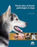
Visual atlas of dental pathologies in dogs
Zubní péče je nezbytná pro prevenci stomatologických problémů u psů, ale některé zubní patologie je někdy opravdu obtížné identifikovat, diagnostikovat a léčit. Kolekcí téměř 400 vysoce kvalitních snímků s přehlednými anatomickými ilustracemi a diagnostickými popisy se podařilo autorovi stvořit z jedinečnou referenční publikaci stomatologie u psů.
Tento atlas představuje nový a cenný příspěvek do literatury veterinární stomatologie. Není učebnicí, spíše využívá široké spektrum dobře uspořádaného obrazového materiálu k jasnému vysvětlení různých patologií u psů."
Španělský autor vybral známé mezinárodní spolupracovníky, aby přispěli do ucelené řady vysoce kvalitního obrazového materiálu zahrnujícího téměř 400 obrázků uspořádaných na více než 100 stranách. Kapitoly a stránky jsou uspořádány v jasném a zajímavém formátu, který efektivně sděluje informace a udržuje čtenářův zájem při cestě za poznáním v oboru veterinární stomatologie. Celková organizace z hlediska etiologie, klinických a diagnostických znaků spolu s dobře označenými diagramy a ilustracemi je významný edukační formát, který umožňuje čtenáři rychle se ponořit do jakékoli požadované úrovně detailů. Zejména srovnání různých vizuálních formátů poskytuje vícerozměrný náhled, například dobře vybrané klinické fotografie a odpovídající rentgenové snímky zdůrazňují důležitou roli radiografie v diagnostice.
Atlas je jak dobře vymyšlený a dobře provedený projekt. Tato kniha má široké pokrytí problematiky zubních patologií u psů, že představuje jedinečný cenný přírůstek do knihovny každého veterinárního lékaře.
Autor: Javier Collados Soto
| Nakladatel | Servet |
|---|---|
| ISBN | 9788416315055 |
| Vydání | 2015 |
| Vazba | tvrdá |
| Počet stran | 124 |
Dental care is essential to prevent dental and oral health problems in dogs, but some dental pathologies are sometimes really difficult to identify, diagnose and treat. With almost 400 high-quality images, clear anatomical illustrations, and diagnostic descriptions, this atlas constitutes a reference handbook for a quick visual identification with a clear orientation towards a precise diagnosis and treatment of the most common dental pathologies.
| 1.Introduction | 1 |
| Dental positional terminology | 2 |
| Histological tooth structure in canines and felines | 3 |
| Diagram of dog and cat dentition | 4 |
| Example of periodontal probing | 6 |
| Periodontal disease classification (AVDC, 2007) | 7 |
| Classification of plaque and dental calculus index (Logan & Boyce, 1994) | 7 |
| Gingival index (Wolf et al., 2005) | 8 |
| Stages of mobility (AVDC, 2007) | 8 |
| Classification of furcation involvement/exposure (AVDC, 2007) | 9 |
| Dental fracture classification (AVDC, 2007) | 10 |
| Endo-periodontal lesions | 12 |
| Classification of tooth resorption (based on the severity of the resorption) (AVDC, 2007) | 13 |
| 2. Oral and dental pathologies in dogs | 16 |
| Dental physiological and radiological oral cavity in dogs | 16 |
| Permanent teeth | 16 |
| Deciduous teeth | 19 |
| Normal tooth eruption | 20 |
| Dog dental radiography models | 21 |
| Dental pathologies | 24 |
| Dental abrasion | 24 |
| Alteration of dental development and eruption | 30 |
| Dental agenesis | 30 |
| Unspecific tooth alterations | 31 |
| Non-physiological diastema | 31 |
| Impacted teeth | 32 |
| Incomplete eruption | 36 |
| Fusion | 38 |
| Gemination | 38 |
| Hyperdontia Dental deviation | 27 |
| Distal deviation | 29 |
| Open bite | 32 |
| Persistent tooth | 33 |
| Dental rotation | 33 |
| Maxillomandibular asymmetry in a side-to-side direction | 34 |
| Systemic pathologies and their consequences in the oral cavity | 35 |
| Leukaemia and feline immunodeficiency (FeLV, FIV) | 35 |
| Pathologies of the oral cavity | 39 |
| Neoplasms | 39 |
| Squamous cell carcinoma | 39 |
| Fibrosarcoma | 44 |
| Metabolic, iatrogenic and other aetiologies | 45 |
| Eosinophilic granuloma | 45 |
| Lentigo | 46 |
| Traumatic aetiology | 47 |
| Soft-tissue lesions. Foreign body | 47 |
| Soft-tissue lesions. Tongue lesions | 47 |
| Soft-tissue lesions. Lesions from occlusion/malocclusion | 49 |
| Soft-tissue lesions. Electric cord burn | 55 |
| Soft-tissue lesions. External trauma | 56 |
| Hard-tissue lesions. Fractures - trauma to the temporomandibular joint area - miscellaneous | 58 |
| Hard-tissue lesions. Mandibular fractures | 60 |
| Hard-tissue lesions. Maxillary fractures | 61 |
| Bacterial, viral, fungal and parasitic origin | 62 |
| Cryptococcosis | 62 |
| Candidiasis | 63 |
| Periodontal disease | 64 |
| Feline gingivostomatitis | 81 |
| Osteomyelitis in the oral cavity | 93 |
| Dental pathologies | 94 |
| Abnormal dental development and eruption | 94 |
| Dental agenesis | 94 |
| Impacted teeth | 95 |
| Gemination | 96 |
| Hyperdontia | (supernumerary teeth) |
| Dental absence | 98 |
| Dental discolouration | 101 |
| Dental fracture | 103 |
| Enamel fracture | 103 |
| Uncomplicated crown fracture | 104 |
| Complicated crown fracture | 105 |
| Complicated crown-root fracture | 112 |
| Root fracture | 116 |
| Tooth resorption | 117 |
| Endodontic disease | 133 |
| References | 139 |
| Alphabetical index | 173 |