Visual atlas of oral pathologies in dogs
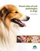
Tento atlas představuje nový a cenný příspěvek ve veterinární stomatologii. Španělský autor si vybral známé mezinárodní spolupracovníky, kteří přispěli k vytvoření komplexního souboru vysoce kvalitního vizuálního materiálu, který obsahuje téměř 500 obrázků uspořádaných na více než 169 stranách. Kapitoly a stránky jsou uspořádány v jasném a zajímavém formátu, který efektivně sděluje informace a udržuje čtenářův zájem na jeho objevné cestě.
Celková organizace z hlediska etiologie, klinických a diagnostických rysů spolu s dobře popsanými diagramy a ilustracemi představuje formát, který čtenáři umožňuje rychle se ponořit do jakékoli požadované úrovně detailů. Zejména juxtapozice různých vizuálních formátů poskytuje vícerozměrný vhled.
Dobrá péče o ústní dutinu je u psů zásadní pro prevenci zdravotních problémů. Některé problémy s ústní dutinou však mohou být někdy opravdu obtížné diagnostikovat a léčit. S téměř 500 vysoce kvalitními obrázky, jasnými anatomickými ilustracemi a podrobnými popisy si tento nový svazek o veterinární stomatologii klade za cíl sloužit jako rychlý vizuální průvodce diagnostikou problémů s ústní dutinou u psů.
Klíčové vlastnosti:
500 vysoce kvalitních obrázků.
Jasné anatomické ilustrace.
Široké pokrytí orálních patologií u psů.
Autor: Javier Collados Soto vykonává stomatologickou činnost a orální chirurgii ve veterinární nemocnici Sierra de Madrid. Působil jako přednášející a koordinátor předmětu Veterinární stomatologie na Fakultě veterinárního lékařství Univerzity Alfonsa X. el Sabia v Madridu.
Autor: Javier Collados Soto
| Nakladatel | Servet |
|---|---|
| ISBN | 9788416315772 |
| Vydání | I. vydání |
| Vazba | pevná |
| Počet stran | 173 |
A good mouth care is paramount to prevent health problems in dogs. However, diagnosing and treating some oral conditions may sometimes be really challenging. With almost 500 high-quality images, clear anatomical illustrations, and thorough descriptions, this new volume on veterinary dentistry aims at being a quick visual guide for treating oral problems in dogs.
| 1. Introduction | 1 |
| • Dental positional terminology | 2 |
| • Histological tooth structure in canines and felines | 3 |
| • Diagram of dog and cat dentition | 4 |
| • Example of periodontal probing | 6 |
| • Periodontal disease classification (AVDC, 2007) | 7 |
| • Classification of plaque and dental calculus index (Logan & Boyce, 1994) | 7 |
| • Gingival index (Wolf et al., 2005) | 8 |
| • Stages of mobility (American Veterinary Dental College, 2007) | 8 |
| • Classification of furcation involvement/exposure (AVDC, 2007) | 9 |
| • Dental fracture classification (AVDC, 2007) | 10 |
| • Endo-periodontal lesions | 12 |
| • Classification of tooth resorption (based on the severity of the resorption) (AVDC, 2007) | 13 |
| 2. Oral pathology in dogs | 15 |
| • Dental physiological and radiological oral cavity in dogs | 16 |
| 1. Permanent teeth | 16 |
| 2. Deciduous teeth | 19 |
| 3. Normal tooth eruption | 20 |
| 4. Dog dental radiography models | 21 |
| • Occlusion and malocclusion | 25 |
| • Occlusion | 25 |
| 1. Incisal area | 25 |
| 2. Area of the first premolars | 25 |
| 3. Canine teeth area | 25 |
| 4. Area of the last premolars and molars | 26 |
| • Malocclusion | 27 |
| 1. Dental crowding | 27 |
| 2. Dental absence | 28 |
| 3. Mandibular distocclusion (class 2 malocclusion) | 30 |
| 4. Mandibular mesiocclusion (class 3 malocclusion) | 35 |
| 5. Tooth displacement | 39 |
| Linguoversion of the mandibular canine teeth | 40 |
| Mesioversion of the maxillary canine teeth | 43 |
| 2. Supernumerary teeth | 44 |
| 3. Maxillary-mandibular asymmetry in a dorsoventral direction (“open bite”) | 47 |
| 4. Rostral crossbite | 48 |
| 5. Caudal crossbite | 49 |
| 6. Persistent teeth | 50 |
| 7. Teeth rotation | 57 |
| 8. Maxillary-mandibular asymmetry in a rostrocaudal direction | 59 |
| 9. Systemic pathologies and their impact on the oral cavity | 60 |
| 10. Secondary hyperparathyroidism | 60 |
| 11. Contact dermatitis | 61 |
| 12. Leishmaniasis | 61 |
| 13. Lymphoma | 63 |
| 14. Systemic lupus erythematosus | 67 |
| 15. Facial paralysis | 67 |
| 16. Bullous pemphigoid | 68 |
| 17. Tetanus | 69 |
| • Pathologies of the oral cavity | 70 |
| • Neoplasms | 70 |
| 1. Non-neoplastic processes. Gingival hyperplasia | 70 |
| 2. Ameloblastoma | 74 |
| 3. Peripheral ameloblastoma (acanthomatous epulis) | 75 |
| 4. Squamous cell carcinoma | 77 |
| 5. Peripheral odontogenic fibroma (fibromatous-ossifying epulis) | 84 |
| 6. Fibrosarcoma | 91 |
| 7. Haemangiosarcoma | 94 |
| 8. Mast cell tumour | 95 |
| 9. Melanoma | 96 |
| 10. Osteosarcoma | 101 |
| 11. Round cell tumour | 102 |
| • Immunological aetiology | 104 |
| Eosinophilic myositis | 104 |
| Mucocutaneous pyoderma | 104 |
| • Congenital and development-related | 105 |
| Juvenile cellulitis | 105 |
| Cleft lip | 106 |
| Macroglossia | 106 |
| Craniomandibular osteopathy | 107 |
| Cleft palate | 108 |
| Cheilitis-dermatitis of the lips | 108 |
| Cleft nose | 109 |
| • Metabolic and iatrogenic aetiology | 109 |
| Calcinosis circumscripta | 109 |
| Oronasal fistula | 110 |
| Eosinophilic granuloma | 111 |
| Salivary sialocoele or ranula | 111 |
| Traumatic aetiology | 111 |
| Soft tissue lesions | 111 |
| Foreign body | 111 |
| • Traumatic dermatitis | 118 |
| • Bucal granuloma (“cheek-chewing lesion”) | 118 |
| • Sublingual granuloma (“tongue-chewing lesion”) | 118 |
| • Lingual lesions | 119 |
| • Lesions from occlusion/malocclusion | 122 |
| • Burns from electric wiring | 123 |
| • Trauma by external bite | 124 |
| Hard tissue lesions | 125 |
| 1. Mandibular fractures | 125 |
| 2. Maxillary fractures | 130 |
| Bacterial aetiology | 134 |
| 1. Periodontal disease | 134 |
| 2. Ulcerative stomatitis. Contact ulcers | 151 |
| 3. Necrotising ulcerative stomatitis | 157 |
| 4. Fistulous tract of dental aetiology | 158 |
| 5. Oronasal fistula | 160 |
| 6. Osteomyelitis in the oral cavity | 163 |
| Viral, mycotic and parasitic aetiology | 167 |
| 1. Demodicosis | 167 |
| 2. Myiasis | 167 |
| 3. Papillomatosis | 168 |
| References | 170 |
| Alphabetical index | 172 |